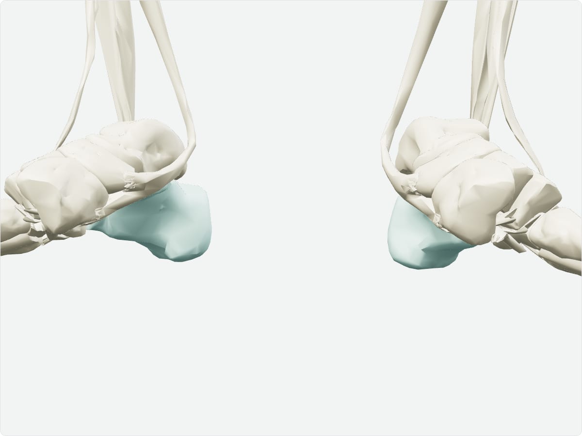
The calcaneus, or heel bone, is the largest tarsal bone of the foot. It forms the prominence of the heel, receives the calcaneal (Achilles) tendon and articulates with the talus above and the cuboid in front. It transmits body weight to the ground.
Key facts
Type
Short (tarsal) bone.
Location
Posterior foot, below the talus.
Landmarks
Calcaneal tuberosity, sustentaculum tali, peroneal trochlea, articular facets for the talus and cuboid.
Articulations
Talus (subtalar joint) and cuboid (calcaneocuboid joint).
Muscle attachments
Gastrocnemius and soleus (via the calcaneal tendon), plantar aponeurosis, short plantar muscles.
3D model: BodyParts3D (CC BY 4.0), adult male reference anatomy. Highlighted structure shown with nearby bones for context.
About the calcaneus
The sustentaculum tali is a shelf that supports the talus and carries the tendon of flexor hallucis longus.
Clinical relevance
Calcaneal fractures usually follow a fall from height and can flatten the arch.
Plantar fasciitis causes heel pain at the origin of the plantar aponeurosis.
Frequently asked questions
What attaches to the calcaneus?
The calcaneal (Achilles) tendon of gastrocnemius and soleus and the plantar aponeurosis.
What is the sustentaculum tali?
A bony shelf on the medial calcaneus that supports the talus.