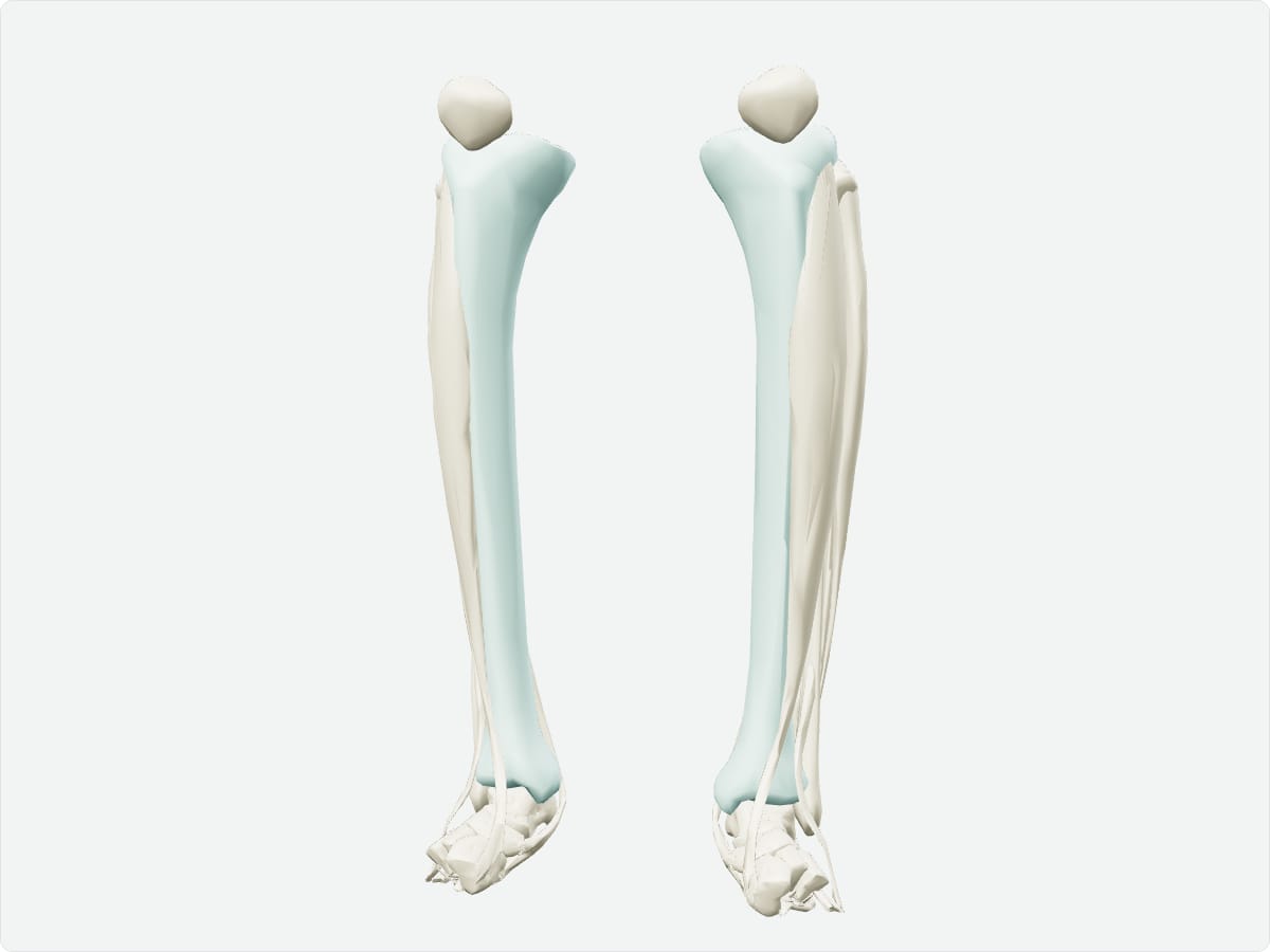
Home / Anatomy / Lower limb / Tibia Quick answer The tibia, or shin bone, is the large weight-bearing bone of the leg. Its proximal condyles articulate with the femur at the knee, and its distal end forms the medial malleolus and the upper surface of the ankle joint with the talus. The anterior border and medial surface lie just under the skin.
Key facts Type Long bone. Location Medial leg. Landmarks Medial and lateral condyles, intercondylar eminence, tibial tuberosity, anterior border, soleal line, medial malleolus, fibular notch. Articulations Femur (knee), fibula (superior and inferior tibiofibular joints), talus (ankle). Muscle attachments Quadriceps via the patellar ligament, sartorius, gracilis, semitendinosus (pes anserinus), semimembranosus, popliteus, soleus, tibialis anterior.
Explore in 3D 3D model: BodyParts3D (CC BY 4.0), adult male reference anatomy. Highlighted structure shown with nearby bones for context.
About the tibia The tibia carries almost all of the body weight through the leg. Its subcutaneous anterior border is easily palpable and makes open fractures common.
Clinical relevance Tibial shaft fractures are the most common long-bone fractures and carry a risk of compartment syndrome. Osgood-Schlatter disease is inflammation at the tibial tuberosity in adolescents. Frequently asked questions Which bone carries most of the weight in the leg? The tibia.
What is the tibial tuberosity? A bony prominence at the top of the shin where the patellar ligament attaches.
Draft: this page has not yet been through editorial review. Read our review process .
Last updated September 30, 2026. Educational use only; not medical advice. Sources are listed on the sources page .