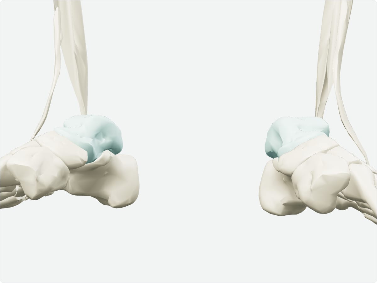
The talus is the tarsal bone that links the leg to the foot. Its dome articulates with the tibia and fibula at the ankle joint and it rests on the calcaneus at the subtalar joint. It has no muscle attachments and is largely covered by articular cartilage.
Key facts
Type
Short (tarsal) bone.
Location
Between the tibia and fibula above and the calcaneus below.
Landmarks
Head, neck, body, trochlea, lateral and posterior processes.
3D model: BodyParts3D (CC BY 4.0), adult male reference anatomy. Highlighted structure shown with nearby bones for context.
About the talus
The wedge shape of the trochlea makes the ankle most stable when the foot is dorsiflexed. Its blood supply enters through the neck, in a retrograde manner.
Clinical relevance
Fractures of the talar neck can disrupt blood supply and cause avascular necrosis of the body.
Frequently asked questions
Does the talus have muscle attachments?
No. It has ligamentous attachments only.
Which joints does the talus form?
The ankle (talocrural), subtalar and talonavicular joints.