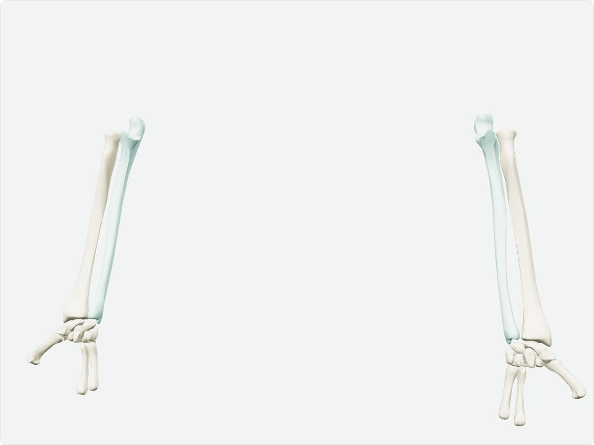
Home / Anatomy / Upper limb / Ulna Ulna: anatomy, landmarks and articulations Quick answer The ulna is the medial bone of the forearm, on the little finger side. Its proximal end forms the hinge of the elbow with the trochlea of the humerus through the trochlear notch between the olecranon and coronoid process. Its distal end is small and articulates with the radius.
Key facts Type Long bone. Location Medial (little finger side) forearm. Landmarks Olecranon, coronoid process, trochlear notch, radial notch, ulnar tuberosity, head, styloid process. Articulations Humerus (elbow), radius (proximal and distal radioulnar joints). Muscle attachments Triceps, brachialis, flexor digitorum profundus, flexor carpi ulnaris, pronator quadratus.
Explore in 3D 3D model: BodyParts3D (CC BY 4.0), adult male reference anatomy. Highlighted structure shown with nearby bones for context.
About the ulna The olecranon forms the point of the elbow. The ulna stays relatively fixed during pronation and supination while the radius rotates around it.
Clinical relevance A direct blow to the forearm can fracture the ulnar shaft (nightstick fracture). An olecranon fracture disrupts triceps function, so the elbow cannot be actively extended. Frequently asked questions What forms the point of the elbow? The olecranon process of the ulna.
Which joints involve the ulna? The elbow joint with the humerus and the proximal and distal radioulnar joints with the radius.
Draft: this page has not yet been through editorial review. Read our review process .
Last updated September 30, 2026. Educational use only; not medical advice. Sources are listed on the sources page .