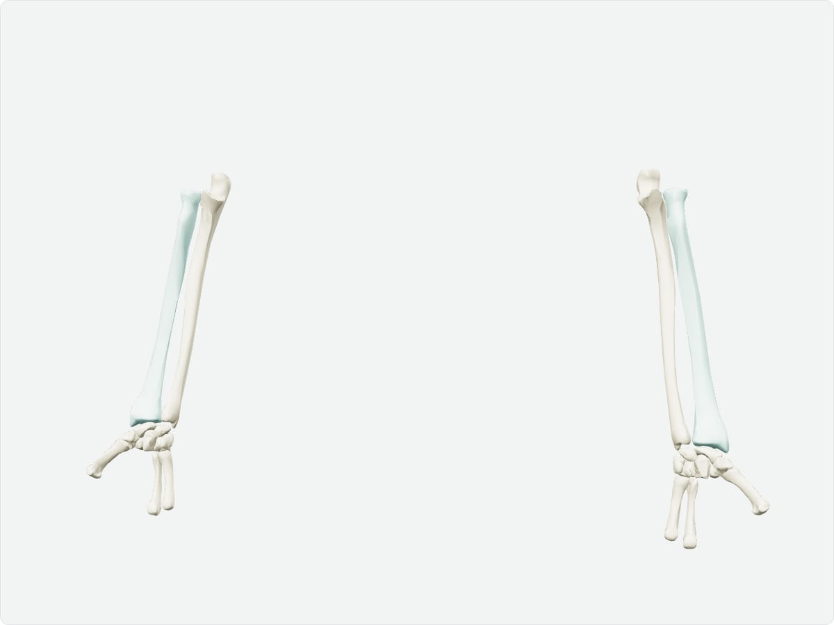
Home / Anatomy / Upper limb / Radius Radius: anatomy, landmarks and articulations Quick answer The radius is the lateral bone of the forearm, on the thumb side. Its head articulates with the humerus and ulna at the elbow and its expanded distal end forms most of the wrist joint with the carpal bones. It rotates around the ulna during pronation and supination.
Key facts Type Long bone. Location Lateral (thumb side) forearm. Landmarks Head, neck, radial tuberosity, styloid process, dorsal (Lister) tubercle, ulnar notch. Articulations Humerus (capitulum), ulna (proximal and distal radioulnar joints), scaphoid and lunate. Muscle attachments Biceps brachii, supinator, pronator teres, pronator quadratus, brachioradialis, flexor digitorum superficialis and others.
Explore in 3D 3D model: BodyParts3D (CC BY 4.0), adult male reference anatomy. Highlighted structure shown with nearby bones for context.
About the radius During pronation the radius crosses over the ulna, pivoting around the proximal and distal radioulnar joints.
Clinical relevance A fall on an outstretched hand commonly fractures the distal radius (Colles fracture) with dorsal displacement. Frequently asked questions What is a Colles fracture? A fracture of the distal radius with dorsal displacement, usually after a fall on an outstretched hand.
Which side of the forearm is the radius on? The lateral (thumb) side in the anatomical position.
Draft: this page has not yet been through editorial review. Read our review process .
Last updated September 30, 2026. Educational use only; not medical advice. Sources are listed on the sources page .