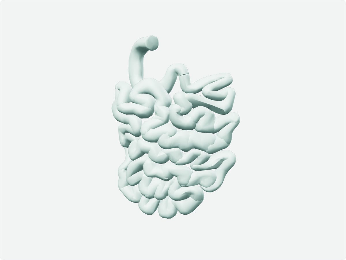
The small intestine is the longest part of the digestive tract, about six metres in living adults, and consists of the duodenum, jejunum and ileum. It completes digestion and absorbs most nutrients through villi on its inner surface. The superior mesenteric artery supplies most of it, and bile and pancreatic enzymes enter at the duodenum.
Key facts
Location
Central abdomen, framed by the large intestine.
Structure
Duodenum (C-shaped, retroperitoneal), jejunum and ileum (suspended by the mesentery); plicae circulares, villi and microvilli.
Function
Final digestion and absorption of nutrients, vitamins and water.
Blood supply
Duodenum: gastroduodenal and superior mesenteric arteries. Jejunum and ileum: superior mesenteric artery.
Innervation
Vagus nerve (parasympathetic) and superior mesenteric plexus (sympathetic).
3D model: BodyParts3D (CC BY 4.0), adult male reference anatomy. Highlighted structure shown with nearby bones for context.
About the small intestine
The atlas models the small intestine as many separate pieces grouped under this concept. Its huge surface area comes from folds, villi and microvilli.
Clinical relevance
Small bowel obstruction is often caused by adhesions after surgery.
Meckel diverticulum is a congenital remnant of the vitelline duct in the ileum.