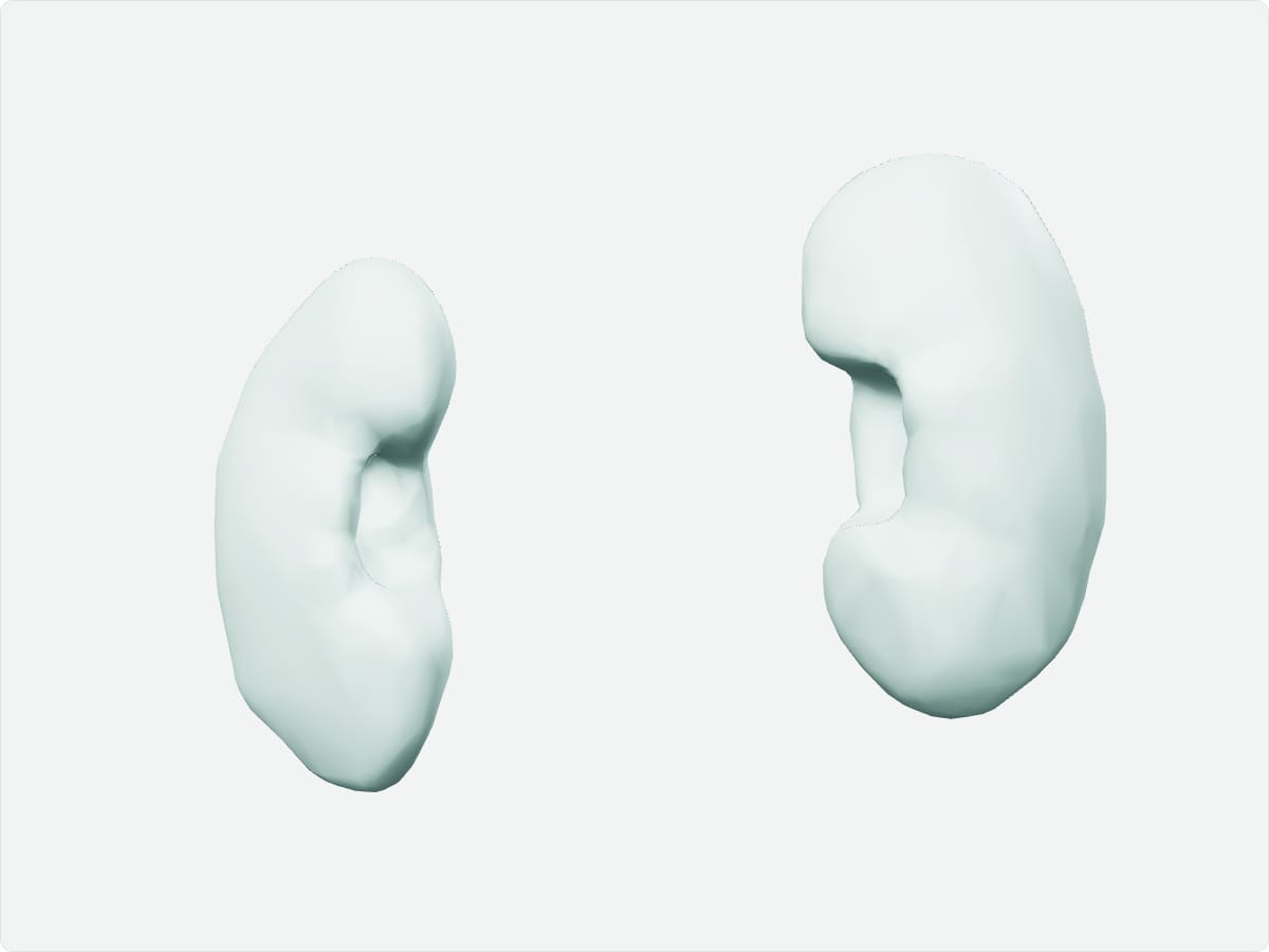
The kidneys are a pair of bean-shaped organs that lie behind the peritoneum on either side of the spine at about T12 to L3. They filter blood, regulate fluid, electrolyte and acid-base balance and produce urine, which drains through the ureters to the bladder. The renal arteries branch directly from the aorta.
Key facts
Location
Retroperitoneal, either side of the vertebral column; the right kidney lies slightly lower than the left because of the liver.
Structure
Renal cortex and medulla, renal pyramids, minor and major calyces, renal pelvis, hilum; nephrons as the functional unit.
Function
Filters blood, regulates fluid and electrolytes, excretes waste, produces erythropoietin and renin, activates vitamin D.
Blood supply
Renal arteries from the abdominal aorta; renal veins drain to the inferior vena cava.
Innervation
Renal plexus, mainly sympathetic (T10-L1).
3D model: BodyParts3D (CC BY 4.0), adult male reference anatomy. Highlighted structure shown with nearby bones for context.
About the kidneys
The kidneys receive about a fifth of cardiac output. The adrenal glands sit on top of each kidney within the same renal fascia.
Clinical relevance
Kidney stones cause colicky pain radiating from the loin to the groin as they pass along the ureter.
Chronic kidney disease reduces the glomerular filtration rate over time.
Frequently asked questions
Where are the kidneys located?
Retroperitoneally on either side of the spine, at roughly the T12 to L3 levels.
What is a nephron?
The functional unit of the kidney, consisting of a glomerulus and a tubule.