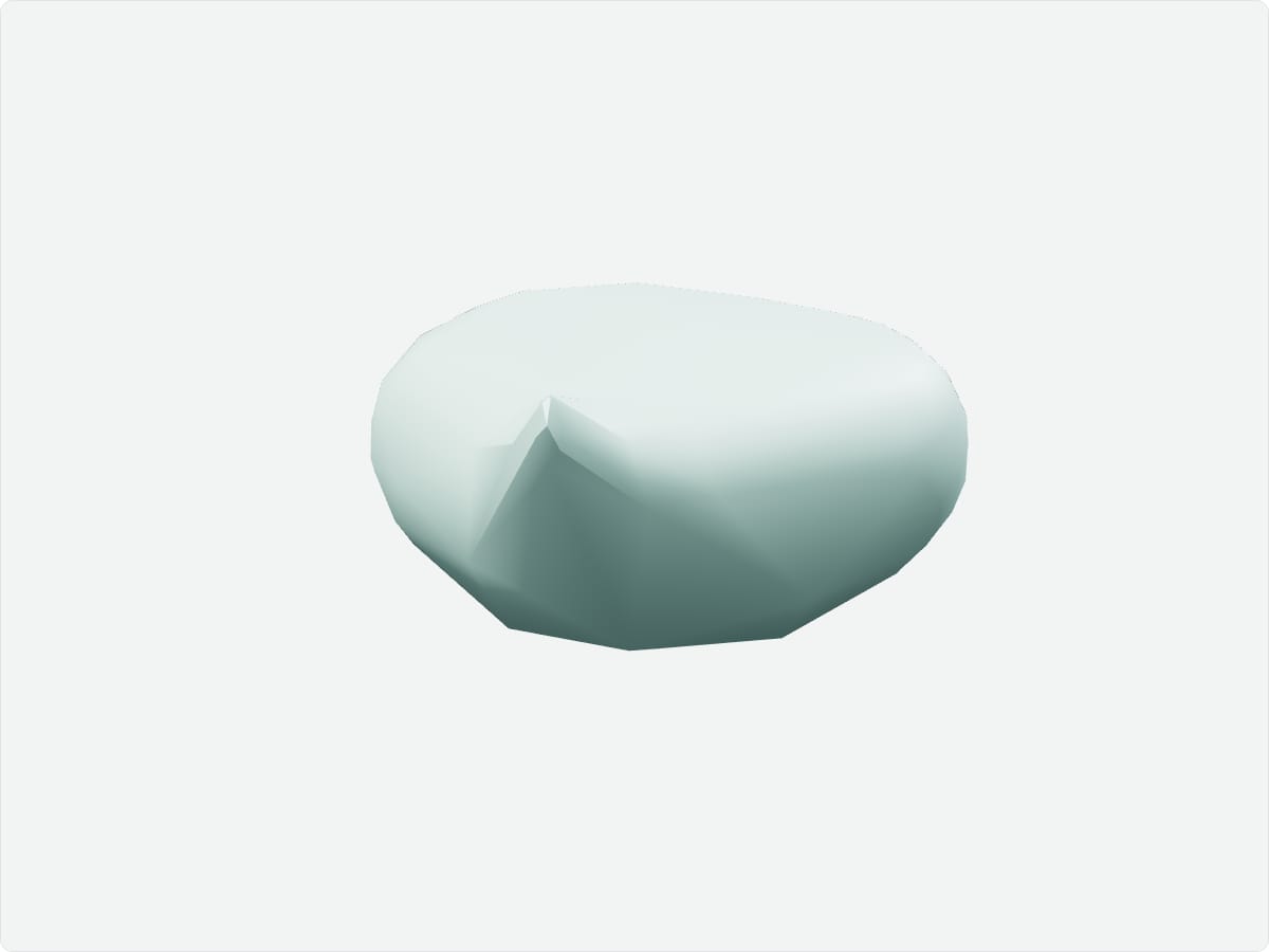
The urinary bladder is a hollow muscular organ in the pelvis that stores urine from the kidneys. Urine enters through the ureters and leaves through the urethra. When full, it rises above the pubic symphysis into the lower abdomen. The detrusor muscle in its wall contracts to empty it.
Key facts
Location
Pelvis, behind the pubic symphysis and in front of the rectum in males.
Structure
Apex, body, fundus and neck; trigone; detrusor muscle; internal and external urethral sphincters.
Function
Stores urine and expels it during micturition.
Blood supply
Superior and inferior vesical arteries from the internal iliac artery.
Innervation
Parasympathetic pelvic splanchnic nerves (S2-S4) for emptying; sympathetic fibres (T11-L2); somatic pudendal nerve to the external sphincter.
3D model: BodyParts3D (CC BY 4.0), adult male reference anatomy. Highlighted structure shown with nearby bones for context.
About the urinary bladder
The bladder can typically hold 400 to 600 millilitres, and the urge to void begins at about 150 to 300 millilitres.
Clinical relevance
A full bladder can be catheterised suprapubically because it rises above the pubic bone.
Urinary tract infections commonly involve the bladder.
Frequently asked questions
What is the trigone of the bladder?
The triangular area between the two ureteric openings and the internal urethral orifice.