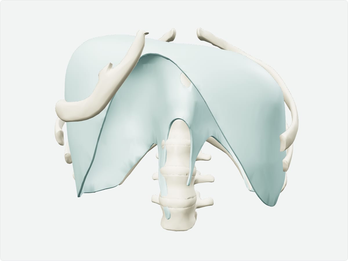
Diaphragm: origin, insertion, nerve supply and action
Also called: thoracic diaphragm
Quick answer
The diaphragm is the dome-shaped muscle separating the chest from the abdomen and is the main muscle of breathing. It arises from the sternum, lower six ribs and lumbar vertebrae and converges on a central tendon. The phrenic nerve (C3-C5) supplies it. Contraction flattens the dome and draws air into the lungs.
Key facts
Origin
Sternal part: back of the xiphoid process. Costal part: inner surfaces of the lower six ribs. Lumbar part: lumbar vertebrae by the crura and arcuate ligaments.
Insertion
Central tendon of the diaphragm.
Innervation
Phrenic nerve (C3-C5) for motor and most sensation.
Blood supply
Pericardiacophrenic, musculophrenic and inferior phrenic arteries.
Action
Main muscle of inspiration: contraction lowers the central tendon and increases thoracic volume.
3D model: BodyParts3D (CC BY 4.0), adult male reference anatomy. Highlighted structure shown with nearby bones for context.
About the diaphragm
Three large openings transmit structures between thorax and abdomen: the vena caval opening at T8, the esophageal hiatus at T10 and the aortic hiatus at T12.
Clinical relevance
A hiatal hernia occurs when part of the stomach protrudes through the esophageal hiatus.
Irritation of the diaphragm can refer pain to the shoulder because the phrenic nerve arises from C3-C5.
Frequently asked questions
Which nerve supplies the diaphragm?
The phrenic nerve, arising from C3, C4 and C5 ("C3, 4, 5 keeps the diaphragm alive").
What passes through the diaphragm?
The inferior vena cava (T8), esophagus and vagus nerves (T10) and the aorta, thoracic duct and azygos vein (T12).