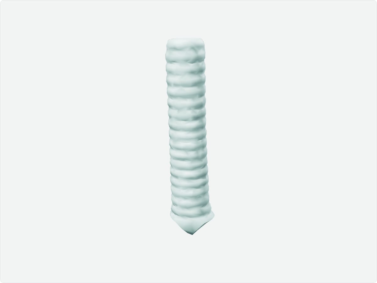
The trachea, or windpipe, is a flexible tube about 10 to 12 centimetres long that conducts air from the larynx to the lungs. It is held open by 16 to 20 C-shaped cartilage rings and divides at the carina into the right and left main bronchi at about the level of T4-T5.
Key facts
Location
From the lower border of the cricoid cartilage at C6, passing through the neck and superior mediastinum.
Structure
C-shaped hyaline cartilage rings, posterior trachealis muscle, ciliated respiratory epithelium; bifurcation at the carina.
Function
Air conduction, warming and humidifying inspired air, and clearing debris by the mucociliary escalator.
Blood supply
Inferior thyroid and bronchial arteries.
Innervation
Vagus and recurrent laryngeal nerves; sympathetic fibres from the sympathetic trunk.
3D model: BodyParts3D (CC BY 4.0), adult male reference anatomy. Highlighted structure shown with nearby bones for context.
About the trachea
The posterior gap in the tracheal rings allows the esophagus to expand during swallowing.
Clinical relevance
Aspirated objects tend to enter the right main bronchus because it is wider, shorter and more vertical.
Tracheostomy creates an airway below the level of the larynx.
Frequently asked questions
Why does the trachea have C-shaped cartilage rings?
The rings keep the airway open, while the gap at the back allows the esophagus to expand during swallowing.
What is the carina?
The ridge at the tracheal bifurcation into the right and left main bronchi.