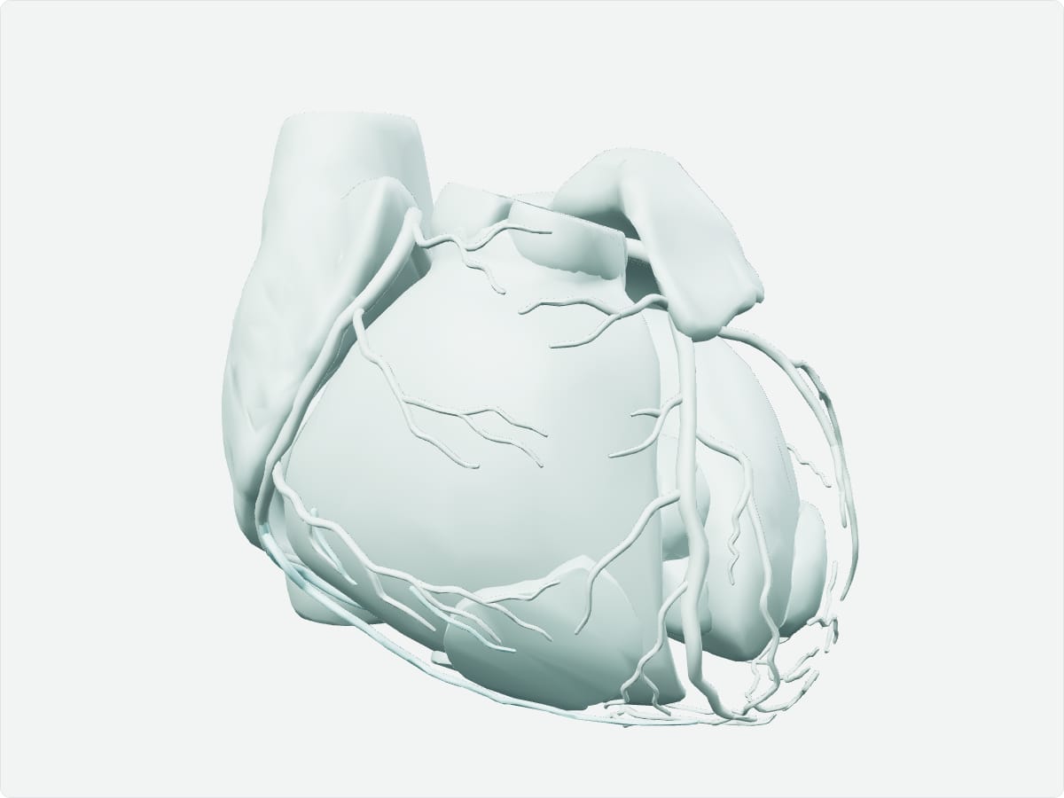
The heart is a four-chambered muscular pump in the middle mediastinum of the chest. The right atrium and ventricle send blood to the lungs, and the left atrium and ventricle send oxygenated blood to the body. Four valves keep blood flowing in one direction, and coronary arteries supply the heart muscle itself.
Key facts
Location
Middle mediastinum, between the lungs, resting on the diaphragm and enclosed in the pericardium.
Structure
Four chambers (right and left atria and ventricles), four valves (tricuspid, pulmonary, mitral and aortic), septa, and the myocardium, endocardium and epicardium.
Function
Pumps blood through the pulmonary and systemic circulations.
Blood supply
Right and left coronary arteries from the ascending aorta; drained mainly by the coronary sinus.
Innervation
Autonomic: sympathetic fibres speed the heart and parasympathetic fibres (vagus) slow it; the sinoatrial node sets the rhythm.
3D model: BodyParts3D (CC BY 4.0), adult male reference anatomy. Highlighted structure shown with nearby bones for context.
About the heart
Each chamber and valve of the atlas model is grouped under the heart concept. The right side of the heart receives deoxygenated blood from the venae cavae, and the left side receives oxygenated blood from the pulmonary veins.
Clinical relevance
Coronary artery disease narrows the arteries that supply the myocardium and can cause angina or myocardial infarction.
Valve disorders such as aortic stenosis or mitral regurgitation are diagnosed by auscultation and echocardiography.
Frequently asked questions
How many chambers does the heart have?
Four: two atria and two ventricles.
What are the heart valves?
The tricuspid, pulmonary, mitral (bicuspid) and aortic valves.