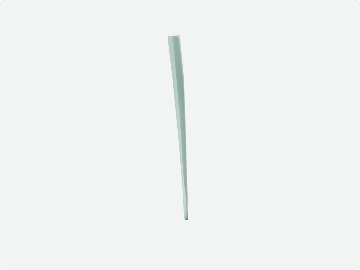
The spinal cord is the cylindrical extension of the central nervous system that runs from the medulla oblongata through the vertebral canal. It ends near L1-L2 in adults at the conus medullaris and carries motor and sensory pathways between the brain and the body through 31 pairs of spinal nerves.
Key facts
Location
Vertebral canal, from the foramen magnum to about the L1-L2 vertebral level.
Structure
Central grey matter surrounding the central canal, surrounded by white matter tracts; cervical and lumbar enlargements; conus medullaris; cauda equina.
Function
Conducts signals between brain and body and mediates spinal reflexes.
Blood supply
One anterior and two posterior spinal arteries reinforced by radicular arteries, including the artery of Adamkiewicz.
Innervation
Gives rise to 31 pairs of spinal nerves: 8 cervical, 12 thoracic, 5 lumbar, 5 sacral and 1 coccygeal.
3D model: BodyParts3D (CC BY 4.0), adult male reference anatomy. Highlighted structure shown with nearby bones for context.
About the spinal cord
The vertebral column grows longer than the spinal cord, so the lower spinal nerve roots descend within the canal as the cauda equina.
Clinical relevance
Lumbar puncture is performed below the level of the conus medullaris, typically at L3-L4, to avoid the cord.
Spinal cord injury causes loss of function below the level of the lesion.
Frequently asked questions
Where does the spinal cord end?
At the conus medullaris, around the L1-L2 vertebral level in adults.