About the cerebellum
Although it holds about half of the brain's neurons, the cerebellum makes up around a tenth of its volume.
Clinical relevance
- Cerebellar lesions cause ataxia, intention tremor and dysmetria on the same side as the lesion.
Also called: little brain
The cerebellum is the large, finely folded structure at the back of the brain below the occipital lobes. It coordinates movement, balance and posture by comparing intended movements with sensory feedback. Damage on one side typically causes ataxia on the same side of the body.
| Location | Posterior cranial fossa, behind the brainstem and beneath the tentorium cerebelli. |
|---|---|
| Structure | Two hemispheres joined by the midline vermis; folia; deep nuclei; three peduncles linking it to the brainstem. |
| Function | Coordination, motor learning, balance and fine control of muscle activity. |
| Blood supply | Superior cerebellar, anterior inferior cerebellar and posterior inferior cerebellar arteries. |
| Innervation | Receives input from the vestibular system, spinal cord and cerebral cortex through the peduncles. |
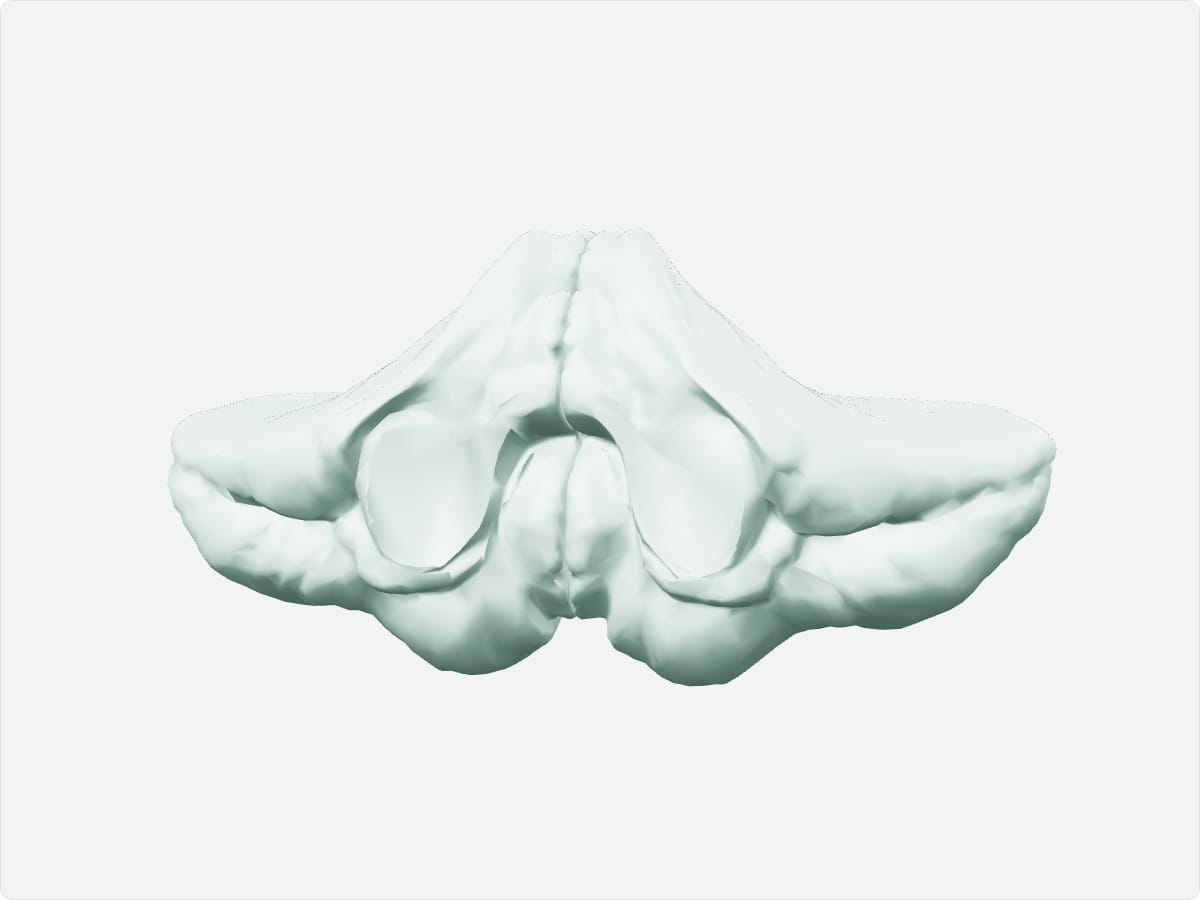
3D model: BodyParts3D (CC BY 4.0), adult male reference anatomy. Highlighted structure shown with nearby bones for context.
Although it holds about half of the brain's neurons, the cerebellum makes up around a tenth of its volume.
It coordinates movement and maintains balance and posture.
The same (ipsilateral) side.
Test yourself
Head and neck structure quiz
Practise origins, insertions, nerve supply and locations with instant feedback.
Explore in context
Open the cerebellum in the full 3D atlas
Toggle body systems, explode the model and inspect every neighbouring structure.