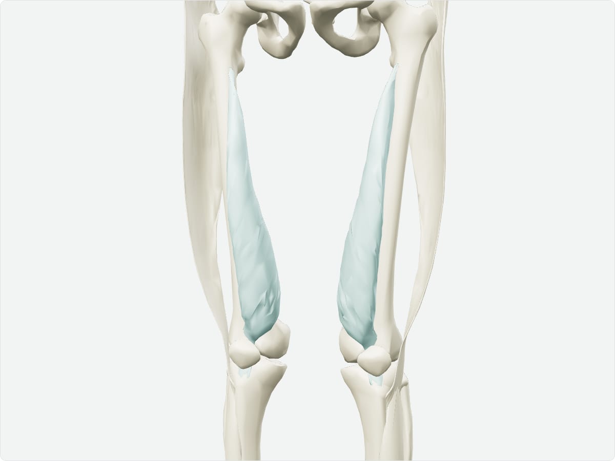
Vastus medialis: origin, insertion, nerve supply and action
Also called: VMO
Quick answer
The vastus medialis forms the medial bulge above the knee. It arises from the intertrochanteric line, the medial lip of the linea aspera and the medial supracondylar line and inserts on the patella and tibial tuberosity through the quadriceps tendon. The femoral nerve supplies it, and its lower oblique fibres keep the patella tracking centrally while extending the knee.
Key facts
Origin
Intertrochanteric line, medial lip of the linea aspera and medial supracondylar line of the femur.
Insertion
Quadriceps tendon and medial border of the patella; tibial tuberosity via the patellar ligament.
Innervation
Femoral nerve (L2-L4).
Blood supply
Femoral artery, descending genicular artery and profunda femoris branches.
Action
Extends the knee; the oblique fibres (vastus medialis oblique) stabilise the patella medially.
3D model: BodyParts3D (CC BY 4.0), adult male reference anatomy. Highlighted structure shown with nearby bones for context.
About the vastus medialis
Weak vastus medialis oblique fibres can allow the patella to track laterally, which is one factor in patellofemoral pain.
Clinical relevance
Strengthening the vastus medialis is a common part of rehabilitation for anterior knee pain.
Frequently asked questions
What is the vastus medialis oblique?
The lowest, obliquely running fibres of the vastus medialis that help keep the patella tracking centrally.