Home / Anatomy / Lower limb / Rectus femoris Rectus femoris: origin, insertion, nerve supply and action Quick answer The rectus femoris is the only quadriceps muscle that crosses the hip. It arises from the anterior inferior iliac spine and the rim of the acetabulum and inserts on the tibial tuberosity through the patella and patellar ligament. The femoral nerve supplies it, and it extends the knee and flexes the hip.
Key facts Origin Straight head: anterior inferior iliac spine. Reflected head: groove above the acetabulum. Insertion Base of the patella, then the tibial tuberosity via the patellar ligament. Innervation Femoral nerve (L2-L4). Blood supply Descending branch of the lateral circumflex femoral artery. Action Extends the knee and flexes the hip.
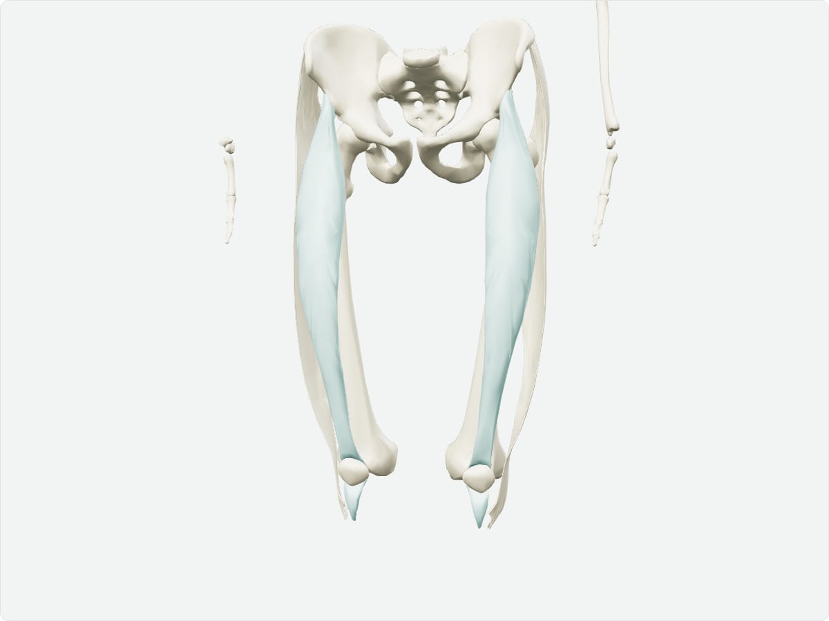
Explore in 3D 3D model: BodyParts3D (CC BY 4.0), adult male reference anatomy. Highlighted structure shown with nearby bones for context.
About the rectus femoris The rectus femoris lies in the middle of the anterior thigh over the vastus intermedius and is bipennate, which gives it considerable strength.
Clinical relevance Rectus femoris strains are common in kicking sports, often at the proximal attachment. The patellar tendon reflex tests spinal segments L3-L4. Frequently asked questions Why is rectus femoris different from the other quadriceps muscles? It is the only one that crosses both the hip and the knee, so it flexes the hip as well as extending the knee.
Where does it originate? From the anterior inferior iliac spine and the superior rim of the acetabulum.
Draft: this page has not yet been through editorial review. Read our review process .
Last updated September 30, 2026. Educational use only; not medical advice. Sources are listed on the sources page .